
J.
Physiol.
(1980),
303,
pp.
391-401
391
With
4
text-fitse
Printed
in
Great
Britain
THE
COURSE
OF
POST-GANGLIONIC
SYMPATHETIC
FIBRES
DISTRIBUTED
WITH
THE
TRIGEMINAL
NERVE
IN
THE
CAT
BY
B.
MATTHEWS
AND
P.
P.
ROBINSON
From
the
Department
of
Physiology
(Oral
Biology),
The
Medical
School,
University
Walk,
Bristol
BS8
1
TD
(Received
14
August
1979)
SUMMARY
1.
The
course
of
post-ganglionic
sympathetic
fibres
to
the
jaws,
face
and
eye
was
investigated
in
cats
by
observing
the
effects
of
nerve
sections
on
responses
evoked
by
stimulation
of
the
cervical
sympathetic
trunk.
2.
Sympathetic
fibres
were
present
in
the
infraorbital
and
inferior
alveolar
nerves.
From
the
superior
cervical
ganglion,
all
of
these
fibres
travelled
in
the
internal
carotid
nerve
and
all
but
a
few
passed
through
the
foramen
lacerum
and
joined
the
trigeminal
nerve
at
its
ganglion.
3.
Compound
action
potentials
were
recorded
from
sympathetic
fibres
in
six
out
of
twenty-seven
teeth.
These
fibres
followed
the
route
described
above.
4.
Sympathetic
fibres
to
the
pupil
and
levator
palpebrae
superioris
passed
from
the
internal
carotid
nerve
to
the
eye
via
the
foramen
lacerum
and
the
superior
orbital
fissure.
Some
fibres
causing
piloerection
in
front
of
the
ear
travelled
by
the
same
route
and
some
travelled
with
the
maxillary
division
of
the
trigeminal
nerve.
5.
Sympathetic
fibres
to
the
nictitating
membrane
followed
a
similar
route
to
those
supplying
the
pupil
except
that
they
entered
the
cranial
vault
through
the
pterygoid
foramen.
6.
The
secretomotor
fibres
to
the
submandibular
salivary
gland
and
some
vaso-
constrictor
fibres
to
the
lip
did
not
travel
with
the
internal
carotid
nerve
or
major
branches
of
the
trigeminal
nerve.
INTRODUCTION
It
is
not
clear
by
what
routes
sympathetic
fibres
travel
from
the
superior
cervical
ganglion
to
supply
the
face
and
jaws
and
it
is
therefore
not
known
whether
section
or
stimulation
of
branches
of
the
trigeminal
nerve
is
likely
to
affect
sympathetic
fibres.
It
is
commonly
taught
that,
in
man,
the
sympathetic
fibres
to
this
region
are
distributed
with
branches
of
the
external
carotid
artery
from
the
external
carotid
plexus
(e.g.
Scott
&
Dixon,
1972).
However,
Langley
(1900)
believed
that
'parts
of
the
skin
and
mucous
membrane
which
receive
their
sole
sensory
supply
from
the
fifth
nerve,
receive
their
sympathetic
supply
also
by
way
of
the
fifth
nerve
..,
and
those
which
run
to
deep
visceral
structures
-
the
salivary
glands
-
accompany
the
arteries.'
It
has
also
been
suggested
(Wilson,
1936;
Christensen,
1940)
that
some
0022-3751/80/8450-0448
$07.50
©
1980
The
Physiological
Society

B.
MATTHEWS
AND
P
P.
ROBINSON
sympathetic
fibres
travel
initially
with
branches
of the
external
carotid
and
then
join
the
terminal
branches
of
the
trigeminal
nerve
(V)
near
their
destination.
Gardner
(1943)
described
branches
from
the
superior
cervical
ganglion
forming
a
plexus
around
the
external
carotid
artery
in
human
cadavers
and
Christensen
(1940)
made
similar
observations
in
cats.
Christensen
also
found
branches
leaving
this
plexus
to
join
the
inferior
alveolar
nerve
and
there
is
physiological
evidence
for
sympathetic
fibres
in
the
supra-orbital
nerve
in
man
(Wilson,
1936)
and
in
the
inferior
alveolar
nerve
in
cat
(Ogilvie,
1969;
Anderson
&
Linden,
1976)
and
dog
(Tonder
&
Naess,
1978).
However,
Taylor
(1950)
found
that,
in
rats,
vasoconstriction
was
produced
in
the
pulps
of
lower
incisors
by
stimulation
of
the
cervical
sympathetic
trunk
but
not
by
stimulation
of
the
inferior
alveolar
nerve.
Langley's
description
receives
support
from
Barlow
&
Root
(1949)
who
demonstrated
fibres
leaving
the
internal
carotid
nerve
to
join
the
inferior
surface
of
the
trigeminal
ganglion
in
the
cat
and
from
Rowbotham
(1939)
who
described
vasodilatation
of
the
face
following
alcohol
injections
into
the
trigeminal
ganglion.
In
preliminary
experiments
in
cats,
it
was
found
that
the
most
reproducible
and
stable
responses
in
the
tissues
innervated
by
the
trigeminal
nerve
that
could
be
evoked
by
sympathetic
stimulation,
were
compound
action
potentials
in
the
inferior
alveolar
and
infraorbital
nerves.
In
the
present
study,
the
course
taken
by
post-
ganglionic
sympathetic
fibres
in
these
nerves
was
determined
by
observing
the
effects
of
nerve
sections
at
different
sites
on
the
compound
action
potentials.
Similar
observations
were
also
made
on
sympathetic
nerves
in
teeth.
The
opportunity
was
also
taken
to
make
some
observations
on
the
effects
of
the
nerve
sections
on
other
sympathetically
mediated
responses
in
the
head
and
neck
region.
A
preliminary
report
of
the
experiments
has
been
published
previously
(Matthews
&
Robinson,
1979).
METHODS
The
experiments
were
carried
out
on
eleven
adult
cats
(2-0-5-0
kg
body
weight);
observations
being
made
on
both
sides
in
nine,
giving
a
total
of
twenty
preparations.
Two
of
the
animals
were
also
the
subjects
of
another
experiment
which
required
the
use
of
a
steroid
anaesthetic
(Saffan,
Glaxo
Laboratories,
induction:
18
mg/kg
I.M.,
maintenance:
2
mg/kg
i.v.).
The
remaining
animals
were
anaesthetized
with
sodium
pentobarbitone
(induction:
42
mg/kg
i.P.,
main-
tenance:
3
mg/kg,
i.v.).
With
both
anaesthetics,
a
maintenance
dose
was
given
whenever
the
flexion
withdrawal
reflex
evoked
by
pinching
the
skin
of
the
foot
returned.
There
was
no
evidence
that
the
choice
of
anaesthetic
influenced
the
results.
Body
temperature
was
maintained
at 37-5
+
0-5
'C
by
an
electric
blanket
controlled
from
a
peritoneal
thermistor.
The
trachea
was
cannulated
and
the
head
stabilized
by
means
of
a
bar
fixed
to
the
frontal
sinus
with
two
self-tapping
screws.
Blood
pressure,
e.c.g.
and
end-tidal
CO2
were
monitored
throughout
each
experiment.
The
cervical
sympathetic
trunk
was
exposed
in
the
neck,
stimulated
electrically
and
the
following
recordings
made:
neurograms
from
the
inferior
alveolar
(in
fifteen
preparations)
and
infraorbital
(two)
nerves
and
the
pulps
of
the
canine
teeth
(ten
upper
and
seventeen
lower),
blood
flow
changes
in
the
lower
lip
(five),
contraction
of
the
nictitating
membrane
(four)
and
levator
palpebrae
superioris
muscle
(four),
dilatation
of
the
pupil
(four),
piloerection
(four),
and
secretion
of
the
submandibular
salivary
gland
(two).
Changes
in
lip
blood
flow
were
also
recorded
during
stimu-
lation
of
the
inferior
alveolar
nerve
(five).
Recordings
from
teeth
were
made
using
the
electrodes
described
by
Horiuchi
&
Matthews
(1974).
Changes
in
lip
blood
flow
were
monitored
by
recording
temperature
changes
with
a
small
Cu-Co
thermocouple
placed
subcutaneously
at
the
muco-cutaneous
junction
of
the
lower
lip
adjacent
to
the
canine
tooth.
Secretion
of
the
submandibular
salivary
gland
was
recorded
by
392

SYMPATHETIC
FIBRES
IN
THE
TRIGEMINAL
NERVE
cannulating
the
submandibular
duct
anterior
to
the
point
where
it is
crossed
by
the
lingual
nerve.
Contraction
of
the
nictitating
membrane
and
levator
palpebrae
superioris
muscle,
dilatation
of
the
pupil
and
piloerection
were
monitored
by
direct
observation.
Stimuli
were
applied
to,
and
recordings
made
from,
nerve
trunks
using
platinum
wire
(diam.
0f
15
mm)
electrodes
under
warm
liquid
paraffin.
The
inferior
alveolar
and
infraorbital
nerves
were
carefully
freed
of
investing
connective
tissue
before
being
placed
on
the
electrodes.
Stimuli
(10
V,
1
0
msec)
were
applied
to
the
cervical
sympathetic
chain
once
a
second.
Neurograms
were
averaged
by
summing
100
successive
records
using
a
PDP
11/10
(Digital
Equipment
Corporation)
computer
with
AR11
interface.
The
preamplifier
bandpass
was
10
Hz-1
kHz.
Fig.
1.
Sites
of
nerve
sections:
(1)
sensory
and
motor
roots
of
trigeminal
nerve
in
posterior
cranial
fossa;
(2)
superior
orbital
fissure;
(3)
foramen
rotundum;
(4)
foramen
ovale;
(5)
foramen
lacerum;
(6)
roof
of
tympanic
bulla;
(7)
mandibular
canal.
To
trace
the
course
of
the
sympathetic
fibres,
the
above
responses
were
observed
before
and
after
section
of
the
following
nerves:
(a)
The
sensory
and
motor
roots
of
the
trigeminal
nerve
posterior
to
the
trigeminal
ganglion
(1
in
Fig.
1).
(b)
The
ophthalmic,
maxillary
or
mandibular
division
of
the
trigeminal
nerve
intracranially
at
the
superior
orbital
fissure,
foramen
rotundum
or
foramen
ovale
respectively
(2,
3
and
4
in
Fig.
1).
All
nerves
passing
through
the
superior
orbital
fissure
were
cut;
no
attempt
was
made
to
identify
them
individually.
(c)
The
internal
carotid
nerve
as
it
passed
through
the
foramen
lacerum
in
the
middle
cranial
fossa
(5
in
Fig.
1).
We
have
confirmed
the
finding
of
Barlow
&
Root
(1949)
that
the
internal
carotid
nerve
crosses
the
roof
of
the
tympanic
bulla
and
enters
the
cranial
vault
between
the
petrous
temporal
and
sphenoid
bones,
i.e.
through
the
foramen
lacerum.
(d)
The
internal
carotid
nerve
in
the
roof
of
the
tympanic
bulla
(6
in
Fig.
1).
In
addition,
neurograms
were
recorded
from
the
lower
canine
pulp
before
and
after
section
of
the
ipsilateral
inferior
alveolar
nerve
(7
in
Fig.
1)
and
contraction
of
the
nictitating
membrane
was
393

B.
MATTHEWS
AND
P.
P.
ROBINSON
observed
before
and
after
intracranial
section
of
the
Vidian
nerve
at
the
pterygoid
foramen,
which
is
anterior
and
medial
to
the
foramen
rotundum
(Jayne,
1898;
Crouch,
1969).
In
those
animals
in
which
intracranial
nerve
sections
were
carried
out,
a
bilateral
decerebration
was
performed
to
gain
access
to
the
nerves.
The
decerebration
was
done
without
clamping
the
carotids.
To
expose
the
contents
of
the
foramen
lacerum
it
was
necessary
to
remove
the
postero-
medial
extension
of
the
alisphenoid,
which
overlies
the
petrous
temporal
bone
and
postero-
lateral
part
of
the
trigeminal
ganglion,
and
to
lift
the
posterior
part
of
the
trigeminal
ganglion.
The
bone
removal
also
exposed
the
greater
superficial
petrosal
nerve,
which
was
divided,
and
improved
access
to
the
sensory
and
motor
roots
of
V.
Section
of
the
internal
carotid
plexus
in
the
tympanic
bulla
was
carried
out
with
the
animal
supine,
after
removing
the
endotympanic
plate
of
the
bulla
(Barlow
&
Root
1949).
In
three
preparations
the
central
end
of
the
inferior
alveolar
nerve
was
stimulated
and
re-
cordings
made
from
the
cervical
sympathetic
trunk.
These
animals
were
paralysed
with
pan-
curonium
bromide
(100
/ag/kg)
and
artificially
ventilated.
Maintenance
doses
of
sodium
pento-
barbitone
were
given
at
the
same
rate
as
that
before
paralysis.
RESULTS
All
the
responses
described
could
only
be
evoked
by
ipsilateral
sympathetic
stimulation,
except
for
some
changes
in
lip
temperature.
Inferior
alveolar
nerve.
In
all
fifteen
preparations
examined,
a
compound
action
potential
was
recorded
from
the
inferior
alveolar
nerve
at
the
level
of
the
mandibular
foramen
during
stimulation
of
the
cervical
sympathetic
trunk.
It
could
be
just
detected
in
single
oscilloscope
sweeps
but
the
results
to
be
described
are
all
based
upon
records
obtained
after
averaging
100
successive
responses
to
improve
the
signal
to
noise
ratio.
The
latency
of
the
compound
action
potential
ranged
from
25
to
88
msec
and
its
duration
was
approximately
150
msec
(Fig.
2).
There
was
no
increase
in
the
amplitude
of
the
response
when
the
stimulus
intensity
was
increased
above
10
V
with
a
duration
of
1
0
msec
(the
parameters
normally
employed)
or
when
the
stimulation
rate
was
reduced
below
once
a
second.
The
response
was
never
affected
by
sectioning
the
sensory
and
motor
roots
of
V
(tested
in
nine
preparations)
(Fig.
2A)
but
was
either
completely
abolished
(one
preparation)
or
almost
completely
abolished
(two
preparations)
(Fig.
2B)
by
intra-
cranial
section
of the
mandibular
division
of
V
at
the
foramen
ovale.
Cutting
the
greater
superficial
petrosal
nerve
(necessary
for
access
to
the
foramen
lacerum)
had
no
effect
whereas
sectioning
the
contents
of
the
foramen
lacerum
either
abolished
the
response
(one
preparation)
or
almost
completely
abolished
it
(three
preparations)
(Fig.
2C).
In
the
latter
three
preparations,
subsequent
sectioning
of
the
mandibular
division
of
V
at
the
foramen
ovale
did
not
affect
the
small
residual
component.
Sectioning
the
internal
carotid
nerve
in
the
bulla
(four
preparations)
always
com-
pletely
abolished
the
response
(Fig.
2D).
Compound
action
potentials
could
always
be
recorded
from
all
four
main
branches
of
the
inferior
alveolar
nerve
below
the
first
premolar
(three
preparations).
Attempts
were
made
to
record
responses
from
the
inferior
alveolar
nerve
during
stimulation
of
the
internal
carotid
nerve
in
the
roof
of
the
bulla.
However,
because
this
nerve
spreads
out
to
form
a
sheet
of
fibres
as
it
passes
through
the
lining
of
the
bulla,
it
was
not
possible
to
isolate
a
sufficient
length
of
nerve
to
place
it
on
stimu-
lating
electrodes,
and
stimulation
in
situ
produced
a
large,
short
latency
response
394

SYMPATHETIC
FIBRES
IN
THE
TRIGEMINAL
NERVE
395
~~~~~~~~~~~~~~~~~~~~~~~~~I
d.
a5
~~~~~~~~~~~~~~~~~~~~~~~~~~~
_111
A~~~~~BL
t
o
0-4
pV
,V
75
msec
25
msec
Fig.
2.
Compound
action
potentials
recorded
from
the
inferior
alveolar
nerve
during
stimulation
of
the
cervical
sympathetic
trunk.
The
signal
was
sampled
at
100,usec
intervals
and
each
record
is
the
average
of
100
successive
responses.
In
each
photograph,
the
lower
trace
represents
the
underlined
segment
of
the
upper
trace
on
an
expanded
time-scale.
The
records
show
the
effects
of
section
of
A,
sensory
and
motor
roots
of
V;
B,
mandibular
division
of
V;
C,
contents
of
foramen
lacerum;
D,
internal
carotid
nerve
in
the
bulla.
The
very
small
residual
components
after
nerve
section
in
B
and
C
were
reproducible
in
repeated
averages.

B.
MATTHEWS
AND
P.
P.
ROBINSON
which
was
attributed
to
stimulus
spread
to
the
roots
of
the
trigeminal
nerve
in
the
overlying
posterior
cranial
fossa.
The
conduction
velocity
of
preganglionic
fibres
was
estimated
(three
preparations)
by
recording
from
the
inferior
alveolar
nerve
during
stimulation
of
the
cervical
sympathetic
trunk
in
two
places
3-4
cm
apart.
The
change
in
the
latency
of
the
inferior
alveolar
nerve
compound
action
potential
indicated
pre-ganglionic
conduc-
tion
velocities
of
6
1,
8-2
and
9
4
m/sec.
Similar
measurements
made
at
two
sites
on
the
inferior
alveolar
nerve
indicated
post-ganglionic
conduction
velocities
(two
preparations)
of
0
9
and
1-3
m/sec.
Stimulation
of
the
central
end
of
the
inferior
alveolar
nerve
(10
V,
1-0
msec)
produced
no
response
in
the
cervical
sympathetic
trunk.
Infra-orbital
nerve.
A
response
very
similar
to
that
in
the
inferior
alveolar
nerve
was
recorded
from
the
infraorbital
nerve
(two
preparations).
It
was
completely
abolished
by
section
of
the
contents
of
the
foramen
lacerum
(one
preparation)
or
the
maxillary
division
of
V
at
the
foramen
rotundum
(one preparation).
Tooth
pulp.
Very
small
amplitude
responses
could
be
recorded
from
six
of
the
twenty-seven
canine
teeth
examined;
two
upper
and
four
lower
(Fig.
3).
Their
latencies
ranged
from
127
to
193
msec.
Neither
upper
nor
lower
(one
preparation
each)
responses
were
affected
by
section
of
the
sensory
and
motor
roots
of
V.
In
the
case
of
the
lower
teeth,
the
response
was
always
abolished
by
section
of
either
the
inferior
alveolar
nerve
alone
(three
preparations)
(Fig.
3A)
or
the
mandibular
division
of
V
alone
(one
preparation).
The
responses
recorded
from
upper
canine
teeth
were
similarly
abolished
by
section
of
either
the
maxillary
division
of
the
trigeminal
nerve
(Fig.
3B)
or
the
internal
carotid
nerve
in
the
bulla
(Fig.
3
C).
In
four
of
the
twenty-
seven
teeth
from
which
recordings
were
made,
dentine
was
exposed
by
fracturing
instead
of
drilling
with
a
dental
bur
(see
Matthews,
1977)
and
compound
action
potentials
were
detected
in
two
of
these.
The
responses
were
similar
to
those
obtained
from
drilled
teeth.
Changes
in
lip
blood
flow.
Sectioning
the
cervical
sympathetic
trunk
(two
preparations)
produced
a
sustained
rise
in
lip
temperature
of
1
0
and
1-7
0C.
Stimulation
of
the
cut
sympathetic
trunk
(five
preparations)
caused
a
marked
drop
in
temperature
of
up
to
3
0C.
Subsequent
section
of
the
inferior
alveolar
nerve
(two
preparations)
resulted
in
a
sustained
drop
in
temperature
of
0-8
and
2-4
'C
but
did
not
affect
the
response
produced
by
sympathetic
stimulation.
Stimulation
of
the
cut
peripheral
end
of
the
inferior
alveolar
nerve
(10
V,
1
0
msec)
produced
either
a
small
rise
or
fall
(two
preparations
each)
in
lip
temperature.
Stimulation
of
the
inferior
alveolar
and
the
sympathetic
trunk
together
produced
a
similar
effect
in
each
animal
to
stimulation
of
the
inferior
alveolar
nerve
alone.
After
the
intravenous
administration
of
the
alpha-blocker
phentolamine
(two
preparations)
in
a
dose
of
2
mg/kg,
there
was
no
change
in
lip
temperature
during
sympathetic
stimulation,
and
inferior
alveolar
nerve
stimulation
produced
a
marked
rise
in
temperature
of
2-3
and
2-5
0C.
Atropine
(100
/tg/kg
i.V.)
given
after
the
phentolamine
did
not
affect
this
response
to
inferior
alveolar
nerve
stimulation.
All
of
these
temperature
changes
were
accompanied
by
contralateral
changes
which
were
much
smaller
but
in
the
same
direction.
Other
responses.
Sympathetic
stimulation
produced
nictitating
membrane
contraction,
levator
palpebrae
superioris
contraction,
pupil
dilation,
piloerection
of
the
fur
between
the
ear
and
the
eye,
and
submandibular
salivary
gland
secretion.
Pupil
dilatation
and
levator
palpebrae
super-
ioris
contraction
were
both
blocked
by
sectioning
the
nerves
passing
through
the
superior
orbital
fissure.
They
were
also
blocked
by
sectioning
the
internal
carotid
nerve
at
the
foramen
lacerum
396

SYMPATHETIC
FIBRES
IN
THE
TRIGEMINAL
NERVE
397
Before
A
004
150
msec
50
msec
002
_
V
75
msec
25
msec
C
,UV
75
msec
25
msec
Fig.
3.
Compound
action
potentials
recorded
from
one
lower
(A)
and
two
upper
(B
and
C)
canine
teeth
during
stimulation
of
the
cervical
sympathetic
trunk.
The
form
of
the
records
is
as
in
Fig.
2
except
that
in
A
the
signal
was
sampled
at
200
#sec
intervals.
The
records
show
the
effects
of
section
of
A,
inferior
alveolar
nerve;
B,
maxillary
division
of
V;
C,
internal
carotid
nerve
in
the
bulla.
B.
MATTHEWS
AND
P.
P.
ROBINSON
or
within
the
tympanic
bulla.
Nictitating
membrane
contraction
was
not
abolished
by
sectioning
the
internal
carotid
nerve
at
the
foramen
lacerum,
however,
and
required
section
of the
Vidian
nerve
or
the
internal
carotid
nerve
in
the
bulla.
It
was
blocked
by
section
of
the
nerves
passing
through
the
superior
orbital
fissure.
Piloerection
of
the
area
between
the
eye
and
the
ear
was
abolished
completely
by
section
of
the
internal
carotid
nerve
in
either
the
bulla
or
the
foramen
lacerum.
It
was
partially
blocked
by
cutting
the
maxillary
division
of
V
and
the
response
disappeared
after
subsequent
section
of
the
nerves
in
the
superior
orbital
fissure.
None
of
the
sections
described
had
any
apparent
effect
on
submandibular
salivary
secretion.
DISCUSSION
Stimulation
of
the
cervical
sympathetic
trunk
produced
compound
action
potentials
in
the
inferior
alveolar
and
infraorbital
nerves.
The
possibility
that
these
responses
were
due
to
some
reflex
pathway
with
an
afferent
limb
in
the
sympathetic
trunk
and
an
efferent
limb
leaving
the
brain
in
the
trigeminal
nerve
was
eliminated
by
demon-
strating
that
they
were
not
abolished
by
section
of
the
sensory
and
motor
roots
of
V.
The
possibility
that
they
were
recorded
from
afferent
fibres
in
the
peripheral
branches
of
the
trigeminal
nerve
which
travelled
back
to
the
central
nervous
system
with
the
sympathetics
was
eliminated
by
demonstrating,
in
the
case
of
the
inferior
alveolar
nerve
response,
that
the
action
potentials
could
not
be
propagated
in
the
reverse
direction
through
the
superior
cervical
ganglion.
It
seems
safe
to
conclude,
therefore,
that
the
responses
were
recorded
from
post-ganglionic
sympathetic
fibres.
Further-
more,
the
results
of
nerve
section
show
that
all
these
fibres
travel
with
the
internal
carotid
nerve
as
far
as
the
tympanic
bulla.
Most
then
enter
the
cranial
cavity
through
the
foramen
lacerum,
still
with
the
internal
carotid
nerve,
and
join
the
trigeminal
nerve
at
its
ganglion.
A
few
of
the
fibres
in
the
inferior
alveolar
nerve
were
not
interrupted
by
section
of
the
nerves
passing
through
the
foramen
ovale
or
the
fora-
men
lacerum
and
can
be
assumed
to
have
crossed
the
base
of
the
skull
outside
the
cranial
vault,
from
the
internal
carotid
nerve
to
the
mandibular
division
of
V.
There
was
no
evidence
of
sympathetic
fibres
leaving
the
external
carotid
plexus
to
join
the
inferior
alveolar
nerve,
contrary
to
the
findings
of
Christensen
(1940).
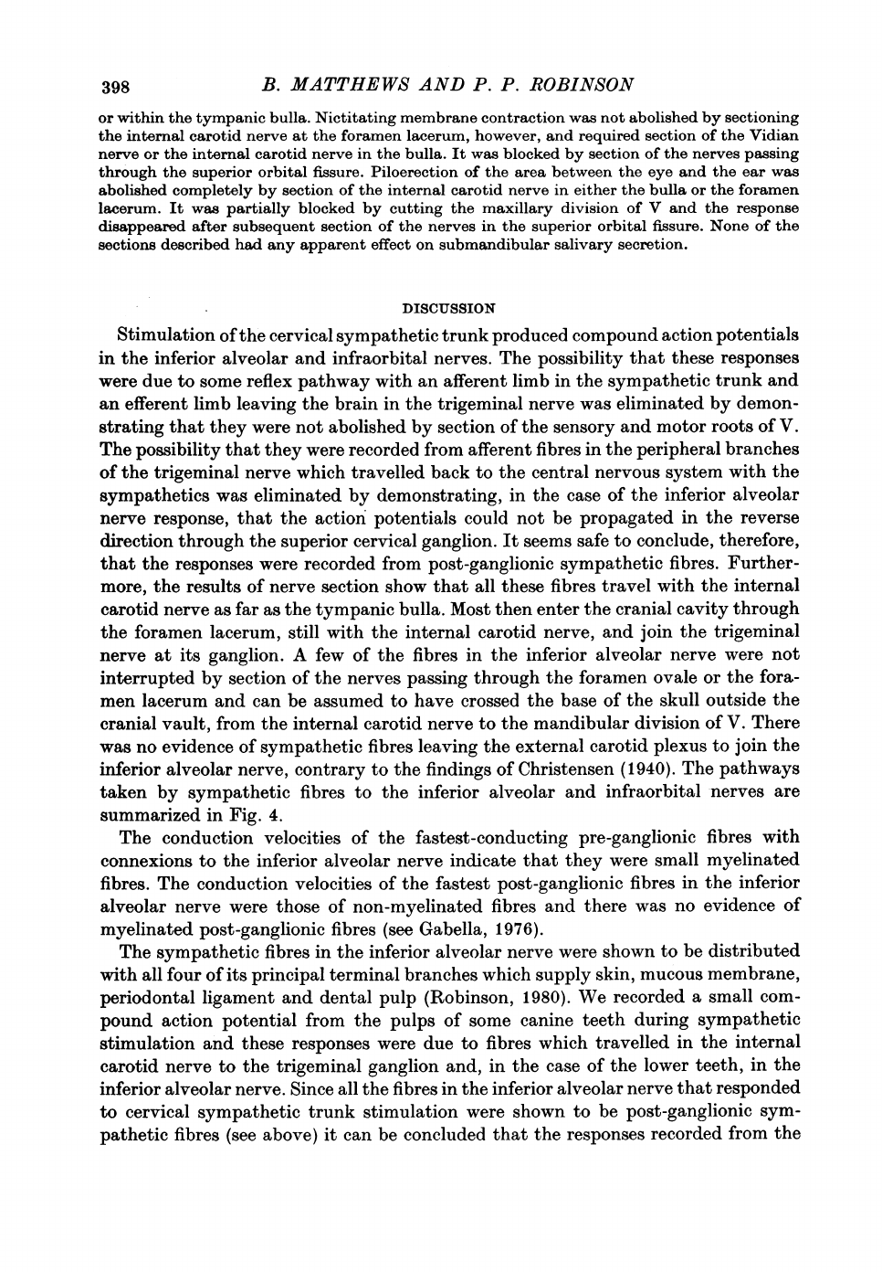
The
pathways
taken
by
sympathetic
fibres
to
the
inferior
alveolar
and
infraorbital
-nerves
are
summarized
in
Fig.
4.
The
conduction
velocities
of
the
fastest-conducting
pre-ganglionic
fibres
with
connexions
to
the
inferior
alveolar
nerve
indicate
that
they
were
small
myelinated
fibres.
The
conduction
velocities
of
the
fastest
post-ganglionic
fibres
in
the
inferior
alveolar
nerve
were
those
of
non-myelinated
fibres
and
there
was
no
evidence
of
myelinated
post-ganglionic
fibres
(see
Gabella,
1976).
The
sympathetic
fibres
in
the
inferior
alveolar
nerve
were
shown
to
be
distributed
with
all
four
of
its
principal
terminal
branches
which
supply
skin,
mucous
membrane,
periodontal
ligament
and
dental
pulp
(Robinson,
1980).
We
recorded
a
small
com-
pound
action
potential
from
the
pulps
of
some
canine
teeth
during
sympathetic
stimulation
and
these
responses
were
due
to
fibres
which
travelled
in
the
internal
carotid
nerve
to
the
trigeminal
ganglion
and,
in
the
case
of
the
lower
teeth,
in
the
inferior
alveolar
nerve.
Since
all
the
fibres
in
the
inferior
alveolar
nerve
that
responded
to
cervical
sympathetic
trunk
stimulation
were
shown
to
be
post-ganglionic
sym-
pathetic
fibres
(see
above)
it
can
be
concluded
that
the
responses
recorded
from
the
398

B.
MATTHEWS
AND
P.
P.
ROBINSON
lower
canine
teeth
were
also
due
to
these
fibres.
The
results
of
nerve
sections
indicate
that
they
reached
the
teeth
via
the
internal
carotid
nerve
and
the
trigeminal
ganglion,
as
described
above
(Fig.
4).
Evidence
for
pulpal
sympathetic
fibres
travelling
by
this
route
was
obtained
by
Ogilvie
(1969)
who
observed
vasoconstriction
in
the
lower
canine
pulp
of
cats
following
stimulation
of
the
internal
carotid
nerve.
He
also
showed
Trigeminal
ganglion
Infra-orbital
nerve~~~~~~~~~~~~~~~~ev
nerve
A_
-------.
-Tympanic
;-~~~~~-;
g < '
I t
~~~~~~~~~~~bulla
teet
carotidd
I
-
-AExternal
Inferior
carotid-
nifeerliloar
~~~~~~artery
alveolar
nerve
Superior
nerve
4 -
~~~~~~~~~~~~~~~~~cervical
\
ganglion
Fig.
4.
The
course
taken
by
sympathetic
fibres
to
the
infra-orbital
and
inferior
alveolar
nerves
and
the
pulps
of
the
canine
teeth.
that
section
of
the
mandibular
division
of
V
intracranially
or
the
inferior
alveolar
nerve
completely
blocked
the
pulpal
vasoconstriction
produced
by
cervical
sym-
pathetic
stimulation.
The
fact
that
compound
action
potentials
could
be
recorded
from
sympathetic
fibres
in
only
six
out
of
twenty-seven
teeth,
and
that
in
these
it
was
of
very
low
amplitude,
indicates
that
the
number
of
sympathetic
fibres
in
the
coronal
pulp
of
cat
canine
teeth
is
small
and
variable.
Rejection
of
the
superior
cervical
ganglion
results
in
degeneration
of
only
a
few
non-myelinated
fibres
in
these
teeth
(Feher,
Csanyi
&
Vajda,
1977).
Christensen
(1940)
also
concluded
that
few
sympathetic
fibres
supplied
the
pulps
of
cat
canine
teeth
but
his
conclusion
was
based
in
part
on
the
incorrect
assumption
that
intracranial
section
of
the
trigeminal
ganglion
would
not
interrupt
any
sympathetic
fibres.
Sympathetic
stimulation
causes
pulpal
vasoconstriction
(e.g.
Edwall
&
Kindlova,
1971;
Scott,
Scheinin,
Karjalainen
&
Edwall,
1972)
and
changes
in
the
response
of
pulpal
afferent
nerve
endings
to
stimulation
(Edwall
&
Scott,
1971;
Matthews,
1976),
although
this
effect
has
not
been
found
in
all
animals
(Matthews,
1976).
399

400
B.
MATTHEWS
AND
P. P.
ROBINSON
The
observations
on
changes
in
lip
temperature
indicate
that
some
sympathetic
vasocon-
strictor
fibres
to
the
lower
lip
travel
with
the
external
carotid
plexus
without
joining
the
inferior
alveolar
nerve
since
section
of
this
nerve
did
not
abolish
vascoconstriction
produced
by
stimu-
lation
of
the
cervical
trunk.
Phentolamine
blocked
this
vasoconstriction
and
there
was
no
evidence
of
vasodilator
fibres
travelling
by
the
same
route.
To
account
for
all
of
the
results
on
lip
blood-flow
it
would
also
be
necessary
to
postulate
that
the
inferior
alveolar
nerve
contained
sympathetic
vasoconstrictor
fibres
and
tonically
active,
non-sympathetic,
atropine-resistant,
vasodilator
fibres.
The
vasodilatation
produced
by
inferior
alveolar
nerve
stimulation
may
have
involved
axon
reflex
mechanisms.
However,
details
of
the
mechanisms
of
these
vascular
effects
requires
further
investigation
using
a
more
accurate
method
for
measuring
skin
and
mucosal
blood
flow.
The
effects
on
lip
blood
flow
reported
here
are
similar
to
changes
in
tooth
pulp
blood
flow
produced
by
sympathetic
trunk
or
inferior
alveolar
nerve
stimulation
(Kroeger,
1968;
Olgart,
Gazelius,
Brodin
&
Nilsson,
1977;
T0nder
&
Naess,
1978).
The
post-ganglionic
sympathetic
fibres
to
the
pupil
and
levator
palpebrae
superioris
muscle
were
shown
to
travel
in
the
internal
carotid
nerve
and
enter
the
cranial
vault
through
the
foramen
lacerum
and
the
orbit
through
the
superior
orbital
fissure.
This
is
in
agreement
with
the
con-
clusions
of
Barlow
&
Root
(1949).
The
course
of
the
fibres
to
the
nictitating
membrane
was
the
same
except
that
they
entered
the
cranial
vault
with
the
Vidian
nerve
through
the
pterygoid
foramen.
Barlow
&
Root
(1949)
suggested
from
their
dissections
that
the
fibres
to
the
nictitat-
ing
membrane
took
the
same
course
as
those
to
the
pupil
and
Thompson
(1961)
reached
the
same
conclusion
in
physiological
experiments,
but
it
seems
that
he
made
no
attempt
to
cut
the
Vidian
nerve
separate
from
the
overlying
trigeminal
ganglion.
Kleijn
&
Socin
(1915)
stated
that
the
fibres
to
the
pupil,
levator
palpebrae
superioris
and
nictitating
membrane
all
travelled
together
through
the
base
of
the
skull
following
a
course
close
to,
but
not
with,
the
Vidian
nerve.
Piloerection
in
the
area
between
the
eye
and
the
ear
was
mediated
partly
by
fibres
travelling
in
the
maxillary
divisions
of
V
and
partly
by
fibres
passing
through
the
superior
orbital
fissure,
presumably
in
the
ophthalmic
division.
These
pathways
are
consistent
with
those
described
by
Langley
(1900).
Secretion
of
the
submandibular
salivary
gland
was
not
abolished
by
section
of
the
internal
carotid
nerve,
supporting
the
conclusions
of
Langley
(1900),
Gardner
(1943)
and
Christensen
(1940)
that
it
receives
its
innervation
direct
from
the
external
carotid
plexus.
The
experiments
were
supported
by
a
grant
from
the
Medical
Research
Council.
P.
P.R.
is
in
receipt
of
a
Medical
Research
Council
Training
Fellowship.
REFERENCES
ANDERSON,
D.
J.
&
LINDEN,
R.
W.
A.
(1977).
Sympathetic
modulation
of
intraoral
mechano-
receptor
activity.
J.
dent.
Res.
56,
D125.
BARLOW,
C.
M.
&
ROOT,
W.
D.
(1949).
The
ocular
sympathetic
path
between
superior
cervical
ganglion
and
the
orbit
in
the
cat.
J.
comp.
Neurol.
91,
195-207.
CHRISTENSEN,
K.
(1940).
Sympathetic
nerve
fibres
in
the
alveolar
nerves
and
nerves
of
the
dental
pulp.
J.
dent.
Res.
19,
227-242.
CROUCH,
J.
E.
(1969).
Text-atlas
of
Cat
Anatomy,
pp.
28-29.
Philadelphia:
Lea
&
Febiger.
EDWALL,
L.
&
KINDLOVA,
M.
(1971).
The
effect
of
sympathetic
nerve
stimulation
on
the
rate
of
disappearance
of
tracers
from
various
oral
tissues.
Acta
odont.
scand.
29,
387-400.
EDWALL,
L.
&
SCOTT,
Jr.,
D.
(1971).
Influence
of
changes
in
micro-circulation
on
the
excitability
of
the
sensory
unit
in
the
tooth
of
the
cat.
Acta
physiol.
scand.
82,
555-566.
FEH1R,
E.,
CSANYI,
K.
&
VAJDA,
J.
(1977).
Ultrastructure
and
degeneration
analysis
of
the
nerve
fibres
of
the
tooth
pulp
in
the
cat.
Archs
oral
Biol.
22,
699-704.
GARDNER,
E.
(1943).
Surgical
anatomy
of
the
external
carotid
plexus.
Archs
Surg.,
Chicago
46,
238-244.
GABELLA,
G.
(1976).
Structure
of
the
Autonomic
Nervous
System,
pp.
40-42.
London:
Chapman
&
Hall.
HORIUCHI,
H.
&
MATTHEWS,
B.
(1974).
Evidence
on
the
origin
of
impulses
recorded
from
dentine
in
the
cat.
J.
Physiol.
243,
797-829.

SYMPATHETIC
FIBRES
IN
THE
TRIGEMINAL
NERVE
JAYNE,
H.
(1898).
Mammalian
Anatomy,
part
1,
p.
283.
Philadelphia:
Lippincott.
KLEIJN,
A.
de
&
SocIN,
C.
(1915).
Zur
ndharen
kenntnis
des
verlaufs
der
postgangliondren
sympathicusbahnen
fur
pupillenerweiterung,
lidspalten6ffnung
und
nickhautretraktion
bie
der
katze.
Pflfgers
Arch.
ge8.
Physiol.
160,
407-415.
KROEGER,
D.
C.
(1968).
Possible
role
of
neurohumoral
substances
in
the
pulp.
In
Biology
of
the
Dental
Pulp
Organ:
A
Symposium,
ed.
FINN,
S.
B.,
pp.
333-346.
Univ.
Alabama:
Univ.
of
Alabama
Press.
LANGLEY,
J.
N.
(1900).
The
sympathetic
and
other
related
systems
of
nerves.
In
Text
Book
of
Physiology,
vol.
2,
ed.
SCHAFER,
E.
A.,
pp.
623-624.
London:
Pentland.
MATTHEWS,
B.
(1976).
Effects
of
sympathetic
stimulation
on
the
response
of
intradental
nerves
to
chemical
stimulation
of
dentine.
In
Advances
in
Pain
Research
and
Theory,
vol.
1,
ed.
BONICA,
J.
J.
&
ALBE-FESSARD,
D.,
pp.
195-203.
New
York:
Raven.
MATTHEWS,
B.
(1977).
Coupling
between
nerve
terminals
in
teeth.
In
Pain
in
the
Trigeminal
Region,
ed.
ANDERSON,
D.
J.
&
MATTHEWS,
B.,
pp.
83-93.
Amsterdam:
Elsevier/North
Holland.
MATTHEWS,
B.
&
ROBINSON,
P.
(1979).
The
course
of
postganglionic
sympathetic
fibres
to
the
face
and
jaws
in
the
cat.
J.
Physiol.
293,
46P.
OGILvI:E,
R.
W.
(1969).
The
vasomotor
innervation
of
the
cat's
lower
right
canine
tooth
pulp.
Anat.
Rec.
163,
237.
OLGART,
L.,
GAZELIUS,
B.,
BRODIN,
E.
&
NIssoN,
G.
(1977).
Release
of
substance
P-like
immuno-
reactivity
from
the dental
pulp.
Acta
physiol.
scand.
101,
510-512.
ROBINSON,
P. P.
(1980).
The
course,
relations
and
distribution
of
the
inferior
alveolar
nerve
and
its
branches
in
the
cat.
Anat.
Rec.
195,
265-272.
ROWBOTHAM,
G.
F.
(1939).
Observations
on
the
effects
of
trigeminal
denervation.
Brain
62,
364-
380.
ScoTT,
J.
H.
DZ&
DixON,
A.
D.
(1972).
Anatomy
for
Students
of
Dentistry,
3rd
edn,
p.
507.
Edin-
burgh:
Churchill
Livingstone.
ScoTT,
Jr.,
D.,
SCHEININ,
A.,
KARJALAINEN,
S.
&
EDWALL,
L.
(1972).
Influence
of
sympathetic
nerve
stimulation
on
flow
velocity
in
pulpal
vessels.
Acta
odont.
sand.
30,
277-287.
TAYLOR,
A.
C.
(1950).
Microscopic
observation
of
the
living
tooth
pulp.
Science,
N.Y.
111,
40.
THOMPSON,
J.
W.
(1961).
The
nerve
supply
to
the
nictitating
membrane
of
the
cat.
J.
Anat.
95,
371-385.
TONDER,
K.
H.
&
NAESS,
G.
(1978).
Nervous
control
of
blood
flow
in
the
dental
pulp
in
dogs
Acta
physiol.
scand.
104,
13-23.
WILSON,
W.
C.
(1936).
Observations
relating
to
the
innervation
of
the
sweat
glands
of
the
face.
Clin.
Sci.
2,
273-286.
401
